Feb 25, 2025

Endobronchial Stenting|A Lifesaving Intervention for Airway Management

Endobronchial Stenting: Restoring Airway Patency with Precision and Innovation

Introduction

Endobronchial stenting has emerged as a pivotal intervention in managing various airway pathologies, particularly those leading to central airway obstruction (CAO). This procedure involves the placement of a stent within the tracheobronchial tree to maintain airway patency, thereby alleviating symptoms and improving the quality of life for affected patients. This comprehensive overview delves into the indications, types, techniques, outcomes, and potential complications associated with endobronchial stenting, with a focus on literature from 2021 to 2025.

image credit


Indications for Endobronchial Stenting

Endobronchial stenting is primarily indicated in scenarios where airway patency is compromised. The main indications include:

  1. Malignant Airway Obstruction: Tumors originating from the trachea or bronchi, or metastatic lesions causing extrinsic compression, can lead to significant airway narrowing. Stenting serves as a palliative measure to relieve obstruction and improve ventilation.

  2. Benign Airway Stenosis: Conditions such as post-intubation tracheal stenosis, inflammatory diseases, or congenital anomalies can result in benign strictures. While surgical resection remains the definitive treatment, stenting offers a less invasive alternative, especially in patients unfit for surgery.

  3. Tracheoesophageal and Bronchopleural Fistulas: Abnormal communications between the trachea and esophagus or pleural space can lead to severe respiratory complications. Stent placement helps seal these fistulas, preventing aspiration and improving respiratory function.

  4. Airway Collapse Due to Extrinsic Compression: Mediastinal masses or lymphadenopathy can exert pressure on the airway, leading to collapse. Stenting provides structural support, counteracting the external compressive forces. 
image credit


Types of Endobronchial Stents

The selection of an appropriate stent is crucial and depends on the underlying pathology, anatomical considerations, and the desired duration of stenting. The primary types include:

1. Silicone Stents

Silicone stents have been widely used in the treatment of both benign and malignant airway obstructions. They are typically inserted using a rigid bronchoscope and provide a non-reactive, biocompatible solution for maintaining airway patency.

Advantages

Biocompatibility: Made of medical-grade silicone, reducing inflammation and tissue reaction.
Easy Removal: Unlike metallic stents, silicone stents can be safely removed, making them ideal for temporary airway management.
Lower Risk of Granulation Tissue Formation: Since they do not embed into the airway wall, the risk of excessive tissue overgrowth is lower.
Customizable: Available in various sizes and shapes; some can be trimmed to fit individual patient anatomy.

Disadvantages

Migration Risk: More prone to displacement, especially in mobile airway regions like the trachea.
Mucus Plugging: Their non-porous surface can accumulate secretions, leading to obstruction.
Rigid Bronchoscopy Requirement: Placement often requires general anesthesia and specialized equipment.
Limited Radial Force: Less effective for extrinsic airway compression compared to metallic stents.

Best Used For

  • Benign airway stenosis (e.g., post-intubation tracheal stenosis).
  • Airway fistula sealing (e.g., tracheoesophageal fistulas).
  • Patients requiring temporary stenting with an option for removal.

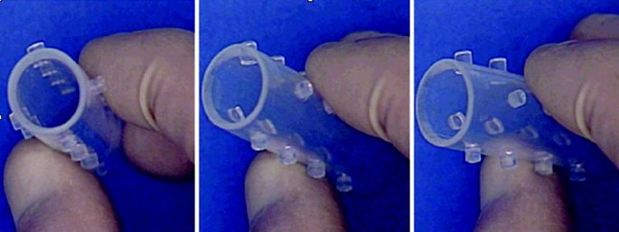
 The original Dumon Stent (Tracheobronxane ®, Novatech, la Ciotat, France)
image credit

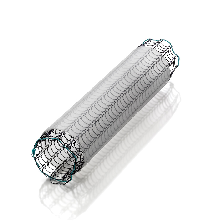
2. Metallic Stents (Self-Expanding and Balloon-Expandable)

Metallic stents, also known as self-expanding metal stents (SEMS), are widely used for malignant airway obstructions due to their excellent radial force and ability to resist external compression. These stents are usually composed of nitinol (nickel-titanium alloy), stainless steel, or cobalt-chromium and are inserted via a flexible bronchoscope.

Types of Metallic Stents

  1. Self-Expanding Metallic Stents (SEMS) – Expand upon deployment, commonly used in malignant obstructions.
  2. Balloon-Expandable Stents – Require external balloon dilation for expansion, used in certain rigid airway obstructions.

Advantages

Strong Radial Force: Ideal for relieving extrinsic compression from tumors or lymphadenopathy.
Minimally Invasive: Can be placed using flexible bronchoscopy under sedation.
Lower Migration Rate: Compared to silicone stents, they stay in place better.
Thin-Walled Structure: Provides a larger airway lumen than silicone stents of the same outer diameter.

Disadvantages

Difficult Removal: Tends to embed into the airway wall, making removal challenging and sometimes requiring surgery.
Granulation Tissue Formation: Chronic irritation leads to tissue overgrowth, potentially causing re-obstruction.
Fracture Risk: Over time, metal fatigue can lead to stent fracture.
Foreign Body Reaction: Some patients may experience chronic inflammation.

Best Used For

  • Malignant airway obstruction (e.g., lung cancer, tracheal tumors).
  • Tracheobronchial collapse due to external compression (e.g., mediastinal masses).
  • Patients with a poor prognosis where stent removal is not anticipated.

image credit

3. Hybrid Stents (Metallic Framework with Silicone Covering)

Hybrid stents combine the benefits of both metallic and silicone stents. They have a metallic framework for radial strength but are coated with silicone to reduce tissue ingrowth and facilitate removal.

Advantages

Lower Granulation Tissue Formation: The silicone covering reduces direct metal-to-tissue contact.
Strong Radial Force: Offers better airway support than pure silicone stents.
Reduced Migration Risk: More stable than traditional silicone stents.
Easier Removal: Can be extracted more easily than bare metallic stents.

Disadvantages

Potential Mucus Plugging: The silicone coating can lead to secretion accumulation.
Limited Flexibility: May not conform as well to highly distorted airways.
Higher Cost: More expensive than conventional silicone or metallic stents.

Best Used For

  • Malignant or benign airway obstruction requiring long-term management.
  • Patients needing a balance between structural support and removability.
  • Cases where granulation tissue formation needs to be minimized.

Comparison Table: Stent Types at a Glance

FeatureSilicone StentsMetallic Stents (SEMS)Hybrid Stents
MaterialMedical-grade siliconeNitinol, stainless steelMetal framework with silicone coating
PlacementRigid bronchoscopyFlexible bronchoscopyFlexible or rigid bronchoscopy
Radial ForceLowHighModerate to high
Migration RiskHighLowModerate
Granulation Tissue FormationLowHighLower than metal stents
Secretion AccumulationHighLowModerate
Ease of RemovalEasyDifficultEasier than metallic stents
Best ForBenign conditions, temporary airway supportMalignant obstruction, external compressionLong-term airway support with removability option

Emerging Trends and Future Developments

The field of airway stenting is evolving, with new technologies aimed at improving stent performance and minimizing complications:

  1. 3D-Printed Stents

    • Custom-made stents tailored to patient-specific airway anatomy.
    • Expected to reduce migration risk and improve patient outcomes.
  2. Biodegradable Stents

    • Designed to degrade over time, eliminating the need for removal.
    • Particularly useful for temporary airway support in benign conditions.
  3. Drug-Eluting Stents

    • Coated with anti-inflammatory or antibiotic agents to reduce granulation tissue formation and infections.
    • May improve long-term outcomes in patients with chronic airway disease.

Techniques of Stent Placement

The placement of an endobronchial stent is a meticulous process that requires careful planning and execution:
  1. Pre-procedural Assessment: Comprehensive evaluation includes imaging studies like computed tomography (CT) scans to delineate the anatomy and extent of obstruction. Pulmonary function tests may also be conducted to assess the baseline respiratory status.

  2. Bronchoscopic Guidance: The procedure is typically performed under general anesthesia using flexible or rigid bronchoscopy. The choice depends on the stent type and the specific clinical scenario.

  3. Fluoroscopic Assistance: Real-time fluoroscopy aids in accurate stent deployment, ensuring proper positioning and expansion.

  4. Post-deployment Evaluation: After placement, bronchoscopy is repeated to confirm stent position and assess for any immediate complications such as bleeding or misplacement.

Outcomes and Efficacy

The success of endobronchial stenting is measured by symptom relief, improvement in airway patency, and quality of life enhancements:

  1. Symptomatic Improvement: Patients often experience immediate relief from dyspnea and other respiratory symptoms post-stenting.

  2. Enhanced Airway Patency: Imaging and bronchoscopic evaluations post-procedure typically show significant improvement in airway diameter.

  3. Quality of Life: Studies have reported improved quality of life scores in patients undergoing stenting for malignant obstructions, with reduced need for additional interventions.ns and Management

While endobronchial stenting is generally safe, potential complications can arise:

  1. Stent Migration: More common with silicone stents, migration can lead to recurrent obstruction or airway injury. Securing the stent adequately during placement and selecting the appropriate size can mitigate this risk.

  2. Granulation Tissue Formation: Particularly associated with metallic stents, excessive tissue growth can re-occlude the airway. Regular bronchoscopic surveillance and, if necessary, interventions like laser therapy can manage this complication.

  3. Infection: The presence of a foreign body can predispose to infections. Prophylactic antibiotics and ensuring optimal stent hygiene are preventive strategies.

  4. Mucus Plugging: Stents can act as a nidus for mucus accumulation, leading to obstruction. Nebulized mucolytics and adequate hydration are recommended to prevent this issue.

Recent Advances and Future Directions

The field of endobronchial stenting is continually evolving:

  1. 3D-Printed Stents: Custom-made stents tailored to individual patient anatomy using 3D printing technology are being explored, promising better fit and reduced complications.

  2. Biodegradable Stents: Research is ongoing into stents that gradually degrade over time, potentially reducing the need for removal procedures and minimizing long-term complications.

  3. Drug-Eluting Stents: Incorporating medications such as anti-inflammatory agents or antibiotics into the stent material aims to prevent complications like granulation tissue formation and infections.

Conclusion

Endobronchial stenting remains a vital tool in the management of various airway pathologies, offering significant symptomatic relief and improved quality of life for patients with airway obstructions. Ongoing research and technological advancements continue to refine stent designs and placement techniques, aiming to enhance efficacy and safety. As with any medical intervention, careful patient selection, meticulous procedural execution, and diligent post-procedural care are paramount to achieving optimal outcomes.

Feb 15, 2025

Brain-Protective Ventilation|Safeguarding the Mind, One Breath at a Time

            Brain-Protective Ventilation

Introduction

Brain-protective ventilation (BPV) is an advanced mechanical ventilation strategy aimed at minimizing secondary brain injury in critically ill patients. This approach balances the need for adequate oxygenation and ventilation while avoiding harmful effects such as increased intracranial pressure (ICP), cerebral hypoxia, and ventilator-induced lung injury (VILI).

Patients with neurological conditions such as traumatic brain injury (TBI), stroke, subarachnoid hemorrhage (SAH), and status epilepticus often require mechanical ventilation. However, conventional ventilation settings may inadvertently worsen brain injury. BPV optimizes lung function while maintaining cerebral homeostasis, making it a crucial strategy in neurocritical care.

This blog provides an in-depth look at brain-protective ventilation, its principles, key strategies, and evidence-based recommendations.

Pathophysiology of Brain-Lung Interaction

The brain and lungs share a complex physiological relationship. Any disturbance in one organ can significantly impact the other. In neurocritical care, improper ventilation can worsen brain injury through the following mechanisms:

  1. Hypoxia and Hyperoxia:

    • Hypoxia (PaO₂ < 60 mmHg) leads to cerebral vasodilation, increasing ICP and worsening brain edema.
    • Hyperoxia (> 150 mmHg) can cause oxidative stress, leading to secondary brain injury.
  2. Hypocapnia and Hypercapnia:

    • Hypocapnia (PaCO₂ < 35 mmHg) induces cerebral vasoconstriction, reducing cerebral blood flow (CBF) and potentially leading to ischemia.
    • Hypercapnia (PaCO₂ > 45 mmHg) causes vasodilation, increasing CBF and ICP, which may lead to brain herniation in severe cases.
  3. Ventilator-Induced Lung Injury (VILI):

    • Lung overdistension, barotrauma, and atelectrauma can release inflammatory mediators, worsening systemic and cerebral inflammation.
  4. Positive End-Expiratory Pressure (PEEP) and Intracranial Pressure:

    • High PEEP increases intrathoracic pressure, reducing venous return from the brain and leading to elevated ICP.

These interactions highlight the importance of BPV strategies to optimize gas exchange without exacerbating brain injury.

Principles of Brain-Protective Ventilation

1. Maintain Adequate Oxygenation (PaO₂ 60–100 mmHg)

  • Target SpO₂ 92–96% to avoid hypoxia and hyperoxia.
  • Use the lowest fraction of inspired oxygen (FiO₂) to prevent oxygen toxicity.

2. Optimize Carbon Dioxide Levels (PaCO₂ 35–45 mmHg)

  • Avoid aggressive hyperventilation, which reduces cerebral perfusion.
  • Permissive hypercapnia should be used cautiously in patients with raised ICP.

3. Use Lung-Protective Ventilation

  • Low tidal volume (Vt): 6–8 mL/kg of predicted body weight to prevent lung overdistension.
  • Plateau pressure < 30 cmH₂O to reduce the risk of VILI.
  • Avoid excessive PEEP, especially in patients with high ICP.

4. Minimize Ventilator-Induced Lung Injury (VILI)

  • Avoid high driving pressures (plateau pressure - PEEP).
  • Use recruitment maneuvers cautiously to prevent derecruitment without increasing ICP.

5. Monitor Intracranial Pressure (ICP) and Cerebral Perfusion Pressure (CPP)

  • Maintain ICP < 20 mmHg and CPP between 60–70 mmHg.
  • Adjust ventilation settings dynamically based on cerebral and pulmonary parameters.

Ventilation Strategies for Specific Neurological Conditions

1. Traumatic Brain Injury (TBI)

  • Ventilation Goal: Maintain PaCO₂ between 35–40 mmHg to prevent ischemia.
  • Oxygenation Strategy: SpO₂ 92–96%, avoiding hyperoxia (> 150 mmHg).
  • PEEP Consideration: Use moderate PEEP (5–8 cmH₂O) cautiously to prevent hypoxia-induced ICP elevation.

2. Stroke (Ischemic & Hemorrhagic)

  • Ventilation Goal: Prevent hypoxia; maintain PaCO₂ in the normal range.
  • Oxygenation Strategy: Avoid hyperoxia, which may worsen reperfusion injury.
  • PEEP Consideration: Avoid excessive PEEP to prevent cerebral venous congestion.

3. Subarachnoid Hemorrhage (SAH)

  • Ventilation Goal: Avoid hypocapnia to prevent vasospasm.
  • Oxygenation Strategy: Prevent hypoxia, which can worsen secondary injury.
  • PEEP Consideration: Minimal PEEP (5–7 cmH₂O) is recommended to optimize lung function.

4. Status Epilepticus

  • Ventilation Goal: Maintain normocapnia to prevent cerebral metabolic crisis.
  • Oxygenation Strategy: Prevent hypoxia to avoid further neuronal damage.
  • PEEP Consideration: Adjust based on lung mechanics while monitoring ICP.

Role of High-Frequency Oscillatory Ventilation (HFOV) in Brain-Protective Ventilation

HFOV is an alternative mode of ventilation in patients with refractory hypoxemia. It provides small tidal volumes at high frequencies, minimizing lung injury.

Potential Benefits in Neurocritical Patients:

  • Reduces lung inflammation and VILI.
  • Prevents excessive fluctuations in ICP by stabilizing intrathoracic pressure.
  • May improve oxygenation without causing cerebral vasodilation.

However, HFOV should be used cautiously, as it may increase mean airway pressure and affect cerebral hemodynamics.

Monitoring and Advanced Ventilation Techniques

1. End-Tidal CO₂ Monitoring (EtCO₂):

  • Provides a non-invasive estimate of PaCO₂ to guide ventilation adjustments.

2. Transcranial Doppler (TCD):

  • Helps assess cerebral blood flow velocity and detect vasospasm.

3. Neuromonitoring:

  • ICP monitoring (via external ventricular drain or intraparenchymal monitor) guides ventilatory adjustments.
  • Brain tissue oxygenation (PbtO₂) measurement optimizes oxygen delivery.

4. Prone Positioning:

  • Used in patients with ARDS to improve oxygenation without excessive PEEP.
  • May benefit neurocritical patients with severe hypoxemia.

5. Extracorporeal Membrane Oxygenation (ECMO):

  • Considered in patients with severe respiratory failure unresponsive to conventional ventilation.
  • Requires careful assessment of its impact on cerebral hemodynamics.

Challenges and Future Directions

Challenges in Brain-Protective Ventilation:

  • Balancing lung protection with optimal cerebral oxygenation.
  • Determining individualized PEEP settings to prevent lung collapse while avoiding increased ICP.
  • Managing ventilator dyssynchrony, which may increase metabolic demand and worsen brain injury.

Future Directions:

  • Personalized Ventilation Strategies:

    • AI-driven ventilator algorithms to optimize settings in real time.
    • Integration of advanced neuromonitoring to tailor ventilation strategies.
  • Novel Respiratory Support Techniques:

    • Development of less invasive oxygenation methods such as high-flow nasal cannula (HFNC) in neurocritical patients.
    • Use of lung-protective ECMO strategies to prevent secondary brain injury.

Conclusion

Brain-protective ventilation is a crucial strategy in the management of neurocritical patients. It requires careful balancing of oxygenation, ventilation, and lung-protective strategies to prevent secondary brain injury.

Key takeaways include:

  • Maintaining adequate oxygenation (PaO₂ 60–100 mmHg) while avoiding hypoxia and hyperoxia.
  • Ensuring PaCO₂ remains within 35–45 mmHg to prevent ischemia or excessive cerebral vasodilation.
  • Using lung-protective ventilation with low tidal volumes and optimal PEEP to prevent VILI.
  • Monitoring ICP and CPP to tailor ventilation settings dynamically.

By integrating these strategies, clinicians can improve outcomes in neurocritical patients, reducing complications and enhancing recovery. As research advances, personalized ventilation approaches will further optimize care for patients requiring brain-protective ventilation.

Jan 25, 2025

Transorbital Intubation|A Rare Airway Frontier

 Transorbital Intubation: A Comprehensive Guide to a Rare Airway Management Technique

Airway management is a cornerstone of critical care, anesthesia, and emergency medicine. While traditional intubation methods such as orotracheal and nasotracheal approaches dominate clinical practice, unusual scenarios occasionally demand unconventional solutions. One such technique, transorbital intubation, is both fascinating and controversial. This guide explores its history, anatomy, indications, procedure, potential risks, and ethical considerations, supported by recent research and clinical experiences.

Understanding Transorbital Intubation

Transorbital intubation refers to establishing an airway through the orbital cavity. It involves creating a passageway via the eye socket and adjacent anatomical structures to access the airway. Although rarely practiced, the procedure has historical significance and remains a topic of intrigue among medical professionals.

This technique is not part of conventional airway management and is typically reserved for extreme, life-threatening scenarios where traditional routes are inaccessible or contraindicated.

image credit


A Historical Overview

The concept of transorbital procedures is not entirely new. Historically, interventions through the orbit were explored in neurosurgery, particularly for lobotomies during the early 20th century. However, the adaptation of this route for airway management emerged as a theoretical discussion rather than a practical application in most settings.

Some wartime anecdotes and rare case reports describe desperate measures where transorbital approaches were considered, but scientific documentation remains limited.

Anatomical Considerations

To understand the feasibility and risks of transorbital intubation, it is crucial to examine the orbital anatomy:

1. Orbital Boundaries

The orbit is a pyramidal cavity housing the eyeball, surrounded by bones like the frontal, zygomatic, ethmoid, and sphenoid.

2. Critical Structures

  • Optic Nerve: Critical for vision and passes through the optic canal.

  • Orbital Arteries and Veins: Highly vascularized, increasing the risk of hemorrhage.

  • Paranasal Sinuses: The ethmoid and sphenoid sinuses are closely associated with the orbit, potentially providing an access route to the airway.

3. Proximity to the Airway

The posterior orbit lies near the nasopharynx and oropharynx, which can theoretically allow a pathway for airway management.


View of the eye cavity (a) tongue, (b) concha. The patient’s maxilla, zygomatic bone, orbita, hard palate, and pterygopalatine fossa were excised image credit

Indications for Transorbital Intubation

While rarely performed, transorbital intubation might be considered in the following extreme situations:

  1. Severe Facial Trauma
    When oropharyngeal and nasopharyngeal access is obstructed due to massive trauma, fractures, or swelling.

  2. Obstruction of Traditional Routes
    Tumors, infections, or foreign bodies blocking conventional airway passages.

  3. Desperate Life-Threatening Emergencies
    Situations where immediate airway access is required, and all other methods fail.

  4. Research and Experimental Settings
    Limited experimental use in cadaveric studies to explore alternative airway pathways.

The Procedure: A Step-by-Step Guide

Disclaimer: This content is for educational purposes only and not intended for clinical application without rigorous training and ethical approval.

1. Preparation

  • Ensure sterile conditions.

  • Administer sedation and analgesia if possible.

  • Use imaging (e.g., CT scan) to assess orbital anatomy and plan the trajectory.

2. Incision and Access

  • Create an incision in the inferior orbital rim or medial canthus, avoiding vital structures like the optic nerve.

3. Passage Creation

  • Carefully navigate through the orbital floor and ethmoid sinus to access the nasopharynx.

  • Use a guidewire or flexible scope to confirm the pathway.

4. Tube Placement

  • Insert the endotracheal tube under direct visualization or with fiber-optic assistance.

5. Securing the Airway

  • Confirm placement using capnography, auscultation, and chest rise.

  • Secure the tube to prevent dislodgement.

Location of the endotracheal tube after intubation


Risks and Complications

Transorbital intubation is fraught with potential complications, making it a last-resort procedure.

  1. Ocular Damage
    Risk of blindness due to optic nerve injury or globe rupture.

  2. Vascular Complications
    Severe hemorrhage from orbital vessels or sinuses.

  3. Infections
    High risk of orbital cellulitis or abscess formation.

  4. Cranial Complications
    Intracranial infections, cerebrospinal fluid leaks, or brain injury.

  5. Failure to Establish Airway
    Inability to secure the airway despite attempts can worsen patient outcomes.

Ethical and Legal Considerations

Given its invasive nature and high risk of complications, transorbital intubation raises significant ethical and legal concerns.

  1. Informed Consent
    In emergencies, obtaining consent may be impossible, but documenting the rationale is crucial.

  2. Training and Expertise
    Only highly skilled practitioners should attempt this procedure, preferably under supervision.

  3. Documentation
    Detailed records of the indication, procedure, and outcomes are essential for legal protection.

Case Studies and Literature Review

Case Study 1: Trauma-Induced Airway Obstruction

A 45-year-old male with severe maxillofacial injuries presented with complete airway obstruction. Traditional intubation methods failed, and a transorbital approach was attempted. The procedure successfully secured the airway, but the patient suffered permanent vision loss.

Literature Review

A 2023 study in the Journal of Emergency Airway Management examined 10 cadaveric models to explore transorbital intubation. The success rate was 40%, highlighting the challenges and risks involved.

The Future of Transorbital Intubation

Advancements in technology and simulation may pave the way for safer approaches to transorbital airway management. Some areas of potential development include:

  1. 3D Imaging and Navigation
    Real-time imaging to enhance precision and reduce complications.

  2. Biocompatible Materials
    Development of specialized tubes or scaffolds designed for transorbital routes.

  3. Training Simulations
    Virtual reality (VR) and cadaveric training to improve practitioner competence.

  4. Research and Innovation
    Studies focusing on anatomical variations and procedural outcomes.

Conclusion

Transorbital intubation, though rarely performed, represents a fascinating blend of anatomical knowledge and clinical ingenuity. While its practical applications remain limited, understanding this technique underscores the importance of adaptability in critical care.

This niche procedure challenges the boundaries of airway management and highlights the resilience and resourcefulness of healthcare providers. However, its risks necessitate extreme caution, thorough training, and ethical vigilance.

Jan 14, 2025

Turning Back Time on Cancer|Korean Scientists Discover Reprogramming Breakthrough

Introduction

The quest to treat cancer effectively and with minimal side effects has taken a monumental leap forward, thanks to Korean scientists. Their groundbreaking discovery enables the reprogramming of cancer cells back into their normal state, potentially redefining the future of oncology. This innovative approach avoids the aggressive destruction of cancer cells, such as in traditional treatments, and instead focuses on reconditioning the cells into a harmless state. Here's a deep dive into this fascinating breakthrough.

image credit


Understanding the Breakthrough

The research team, led by Professor Kwang-Hyun Cho from the Department of Bio and Brain Engineering at KAIST (Korea Advanced Institute of Science and Technology), has introduced a transformative approach to cancer treatment. By leveraging a "digital twin" of the gene network associated with cellular differentiation, they identified key molecular switches that guide cancer cells back to their normal functions.

These switches, when activated, influence cancer cells to follow a path of differentiation similar to normal cells. The approach was validated through extensive molecular and cellular experiments, as well as animal studies. This method doesn’t rely on destroying cancer cells—a strategy that often leads to severe side effects—but instead “unlocks” the cells’ potential to return to a healthy state.

The Science Behind the Discovery

This revolutionary therapy rests on the understanding that cancer cells often represent an arrested state of differentiation, where they lose their specialized functions and proliferate uncontrollably. By analyzing the differentiation trajectories of normal cells, the Korean research team identified pivotal transcription factors—proteins that regulate gene expression—which can guide cancer cells toward reprogramming.

Using computational simulations and advanced gene-editing technologies such as CRISPR, the researchers applied these molecular switches to colon cancer cells. Astonishingly, the cells began to exhibit normal growth patterns and regained functional characteristics akin to their healthy counterparts. These findings were further corroborated through animal model experiments, which demonstrated the reduction of tumor sizes without adverse effects.

Implications for Oncology

The ability to reverse cancer cells offers numerous advantages over traditional cancer treatments:

  1. Minimal Side Effects: Unlike chemotherapy or radiation, this approach avoids damage to healthy cells and tissues.

  2. Reduced Risk of Recurrence: By addressing the fundamental properties of cancer cells, this treatment minimizes the chance of resistance and relapse.

  3. Potential for Broad Application: While initial experiments focused on colon cancer, this method may be adaptable to various cancer types.

Overcoming Challenges

Despite its promise, the path to clinical implementation faces several challenges:

  1. Comprehensive Testing: Extensive clinical trials are required to validate the therapy’s safety and efficacy in humans.

  2. Ethical Considerations: The use of gene-editing tools like CRISPR necessitates thorough ethical scrutiny to prevent misuse.

  3. Scalability: Adapting this personalized therapy for widespread application will require significant technological advancements.

Conclusion

The discovery by Korean scientists marks a significant milestone in the fight against cancer, shifting the paradigm from aggressive eradication to controlled reprogramming. By focusing on restoring normal cellular functions, this approach provides a promising alternative to traditional treatments, with fewer side effects and a reduced risk of recurrence. While challenges remain, this innovative method lays the groundwork for a new era in oncology, offering hope to millions of patients worldwide.

Jan 13, 2025

Facing the Challenge|Prone Ventilation as the Game-Changer for ARDS Patients

Introduction

 Prone ventilation, or placing patients face-down, has emerged as a pivotal intervention in managing Acute Respiratory Distress Syndrome (ARDS). This technique has garnered significant attention between 2020 and 2024, especially during the COVID-19 pandemic, highlighting its relevance for respiratory therapists and physicians.

Understanding ARDS

ARDS is a severe lung condition characterized by rapid-onset inflammation and increased pulmonary vascular permeability, leading to hypoxemia and respiratory failure. Common causes include pneumonia, sepsis, trauma, and inhalation injuries. The hallmark of ARDS is the accumulation of fluid in the alveoli, resulting in impaired gas exchange and decreased lung compliance.

Image credit


The Berlin Definition


The Rationale Behind Prone Ventilation

The supine position in mechanically ventilated patients can exacerbate lung injury due to gravitational forces, leading to atelectasis in dorsal lung regions and overdistension in ventral areas. Prone positioning redistributes these forces, promoting more uniform lung aeration and perfusion. This results in improved oxygenation and reduced ventilator-induced lung injury (VILI).

image credit


Physiological Benefits of Prone Positioning

1. Improved Oxygenation: Prone positioning enhances ventilation-perfusion matching by recruiting posterior lung regions, leading to better oxygenation. Studies have shown significant improvements in the PaO₂/FiO₂ ratio with prone positioning. 

2. Reduction in VILI: By promoting more homogeneous lung inflation, prone positioning decreases the risk of VILI, a critical consideration in ARDS management.

3. Enhanced Secretion Clearance: The prone position facilitates mucus drainage, reducing the risk of ventilator-associated pneumonia.

Understanding West Zones

The lungs are divided into three zones based on the relationship between alveolar pressure (PA), arterial pressure (Pa), and venous pressure (Pv):

  1. Zone 1 (Dead Space): PA > Pa > Pv

    • Minimal or no blood flow due to high alveolar pressure compressing capillaries.
    • Typically occurs in the apex of the lungs in the upright position.
  2. Zone 2 (Waterfall Zone): Pa > PA > Pv

    • Blood flow depends on the difference between arterial and alveolar pressures.
    • Middle regions of the lungs in upright patients.
  3. Zone 3 (Perfusion Zone): Pa > Pv > PA

    • Continuous blood flow occurs as capillaries are fully distended.
    • Typically found in the dependent regions of the lungs (posterior in supine, anterior in prone).
    • image credit


West Zones in Prone Ventilation

When a patient is in the prone position, the distribution of West Zones shifts due to the reorientation of the lung and chest wall relative to gravity:

  • Supine Position:

    • The dorsal (posterior) lung regions, typically Zone 3, are compressed by the heart and abdominal contents.
    • Ventilation predominantly occurs in non-dependent ventral (anterior) regions, leading to ventilation-perfusion (V/Q) mismatch.
  • Prone Position:

    • Dorsal regions, previously compressed, now expand as gravitational and compressive forces redistribute.
    • Ventilation increases in these dorsal regions, which often correspond to areas with better perfusion (Zone 3).
    • This leads to improved V/Q matching and oxygenation

Clinical Evidence Supporting Prone Ventilation (2020-2024)

Recent studies have reinforced the efficacy of prone ventilation in ARDS patients:

Mortality Reduction: Prone ventilation has been linked to a reduction in mortality among ARDS patients. Research indicates that employing prone positioning for at least 16 hours daily can significantly decrease 90-day mortality without substantial adverse effects. 

Synergistic Effects with Low Tidal Volume Ventilation: When combined with low tidal volume ventilation, prone positioning may exhibit synergistic lung-protective effects. The survival advantage of the prone position seems contingent on the concurrent use of low tidal volumes. 

Feasibility and Safety: Extended prone position ventilation has been deemed feasible and relatively safe for treating critically ill patients with ARDS, including those with COVID-19-related ARDS. This suggests potential implications for a broader acceptance of prone ventilation in ARDS management. 

Implementation Strategies for Respiratory Therapists and Physicians

1. Patient Selection: Not all ARDS patients are ideal candidates for prone ventilation. Consider factors such as hemodynamic stability, absence of contraindications (e.g., spinal injuries), and the severity of hypoxemia.

2. Timing and Duration: Early initiation of prone positioning, especially within the first 48 hours of ARDS diagnosis, is associated with better outcomes. A cumulative duration of more than 32 hours during the first 48 hours of ICU admission has been linked to improved outcomes. 

3. Team Coordination: Prone positioning requires a coordinated multidisciplinary approach to ensure patient safety and minimize complications. Regular training and simulation exercises can enhance team efficiency.

4. Monitoring and Assessment: Continuous monitoring of oxygenation parameters, hemodynamics, and potential pressure points is crucial. Adjustments should be made based on the patient's response to prone positioning.

image credit


Challenges and Considerations

While prone ventilation offers significant benefits, it also presents challenges:

Complications: Potential complications include pressure injuries, accidental extubation, and facial edema. Implementing evidence-based strategies to prevent these complications is crucial. 

Resource Intensiveness: Prone positioning is labor-intensive and requires adequate staffing and equipment. Ensuring the availability of trained personnel and appropriate equipment is essential for successful implementation.

Patient Tolerance: Not all patients may tolerate prone positioning. Regular assessment and prompt management of any adverse effects are necessary to ensure patient comfort and safety.

Conclusion

Prone ventilation has solidified its role as a cornerstone in the management of ARDS, offering improved oxygenation and survival benefits. The period from 2020 to 2024 has provided robust evidence supporting its efficacy, particularly during the challenges posed by the COVID-19 pandemic. For respiratory therapists and physicians, understanding the nuances of prone positioning, from patient selection to implementation and monitoring, is essential. By embracing this technique, healthcare professionals can enhance patient outcomes in ARDS, turning the tide in the battle against this formidable condition.

Jan 10, 2025

The Heimlich Maneuver: A Lifesaving Technique for Choking Emergencies

INTRODUCTION 

The Heimlich maneuver was initially introduced in 1974 by Dr. Henry Heimlich after proving his theory that the reserve of air in the lung could serve to dislodge objects from the esophagus by quick upwards thrust under the ribcage. 

Abdominal thrusts or the Heimlich maneuver is a first-aid procedure used to treat upper airway obstruction caused by a foreign body. This skill is commonly taught during basic life support (BLS) and advanced cardiac life support (ACLS) classes. The abdominal thrust maneuver can be performed in both children and adults via different techniques

Image credit

ANATOMICAL AND PHYSIOLOGICAL CHANGES 

Anatomicaly ,Foreign objects associated with choking are usually stuck above the cricoid cartilage in the supra-laryngeal area. As for the maneuver itself, the thrusts must be executed over the epigastric region just below the ribcage and directed upwards towards the head of the patient.

Physiologically, the abdominal thrust maneuver is effective due to increasing intrathoracic pressure affecting the lung/airway, stomach, and esophagus produced by diaphragmatic thrusts.

INDICATION 

 - Conscious choking victims 

 - Subjects with universal choking signs mainly consists of inability to speak, breathe, or cough   while holding both hands up to one's own throat. 

Image credit


CONTRAINDICATIONS 

 - No absolute contraindication

 - But ,the Maneuver is not recommended by AHA for infants and unconscious patients 

TECHNIQUE 

1) PREPARATION :Cases  of choking happen in mere seconds and unexpectedly, making preparation nearly impossible. As mentioned above, the Heimlich maneuver is taught during BLS for the conscious choking adult.

2) HOW TO PERFORM:

  - It is performed by a bystander on a person who appears to be choking. 

  - The bystander stands behind the subject and wraps his/her arms around the upper abdominal region, about two inches above the belly button. 

  - Making a fist with one hand and wrapping the other hand tightly over the fist and delivering five sharp midline thrusts inward and upward.

  - Recently, other techniques such as the circumferential (horizontal) abdominal thrust, chair thrust, and auto up-thrust have been studied comparing the gastric and esophageal pressures generated with each, finding that chair thrusts might be more effective in these parameters.

                                                                       Image Credit

COMPLICATIONS 

   - Displacement of diaphragm 

   - sudden intrathoracic pressure increases 

   - Rib fracture 

   - Gastric or esophageal perforation

CONCLUSION

The Heimlich maneuver, introduced by Dr. Henry Heimlich in 1974, remains an essential life-saving technique for managing upper airway obstructions caused by foreign bodies. Its simplicity, effectiveness, and widespread teaching in BLS and ACLS courses have made it a cornerstone of first-aid practices. By understanding its anatomical and physiological basis, proper indications, and technique, responders can effectively perform the maneuver to prevent fatalities from choking incidents. However, awareness of potential complications and contraindications is vital for ensuring patient safety. Continuous education and adaptation of newer techniques, like chair thrusts, further enhance the efficacy of this life-saving intervention.

Endobronchial Stenting|A Lifesaving Intervention for Airway Management

Endobronchial Stenting: Restoring Airway Patency with Precision and Innovation Introduction Endobronchial stenting has emerged as a pivotal ...