Dec 27, 2024

Silent Pulse: Unveiling the Mystery of Takayasu's Arteritis

Introduction 

Imagine being in the prime of your life and suddenly experiencing tiredness, diminished strength, and a pulse that seemingly vanish. This is a daily reality for people who have Takayasu's arteritis, not just a hypothetical situation. Despite the difficulties, people can live happy, fulfilled lives if they recognize the disease and use effective treatments.
Image credit


Understanding Takayasu’s Arteritis

 Takayasu's arteritis (TA) is a rare chronic inflammatory disease that affects large blood arteries, mainly the aorta and its branches. Although it can happen to anybody, it generally affects young women, particularly those of Asian heritage. It often referred to as the "pulse-less disease" because of its defining characteristic, which is weak or absence of pulses.

The Enigma of Its Cause

 The cause of Takayasu's arteritis is uncertain. But according to researchers, it's an autoimmune reaction in which the body's arteries are wrongly attacked by the immune system. Its onset may also be influenced by environmental stimuli and genetic variables.
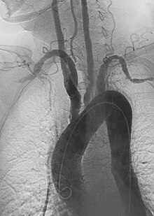
Left anterior oblique  angiographic image of Takayasu's arteritis showing areas of stenosis in multiple great vessels Image Credit


Recognizing the Symptoms

Early symptoms of TA are often vague, making diagnosis a challenge. These may include:

  • Fatigue

  • Fever

  • Unexplained weight loss

  • Joint pain

As the condition progresses, reduced blood flow leads to more distinct symptoms:

  • Weak or absent pulses

  • High blood pressure

  • Pain or cramping in arms or legs during activity

  • Dizziness or fainting

  • Visual disturbances

Diagnosing Takayasu’s Arteritis

Given its rarity, diagnosing TA requires thorough investigation. Physicians typically use:

  • Imaging Tests: MRI, CT scans, or angiography to identify arterial inflammation or narrowing.

  • Blood Tests: To detect inflammatory markers like C-reactive protein (CRP) or erythrocyte sedimentation rate (ESR).

  • Image Credit


Treatment Options

While there is no cure for Takayasu’s arteritis, treatments aim to manage inflammation and prevent complications. Key approaches include:

  • Corticosteroids: These provide rapid relief from inflammation.

  • Immunosuppressants: Medications like methotrexate or azathioprine help sustain long-term control.

  • Biologic Therapies: Drugs such as tocilizumab target specific inflammatory pathways for better management.

  • Surgical Solutions: In cases of severe arterial damage, procedures like bypass surgery or stent placement may be necessary.

Thriving with Takayasu’s Arteritis

Adapting to life with TA involves embracing new routines and priorities. Here are strategies to thrive:

  1. Educate Yourself: To speak up for your health and make educated decisions, educate yourself about the disease you have.

  2. Seek Support: Join support groups or online communities to connect with others who understand your journey.

  3. Practice Self-Care: Focus on adequate rest, balanced nutrition, and appropriate physical activity to boost your overall well-being.

  4. Monitor Regularly: Keep track of your blood pressure and schedule routine medical check-ups.

Conclusion

Living with Takayasu’s arteritis poses unique challenges, but it also fosters resilience and adaptability. By staying informed, building a support network, and working closely with healthcare professionals, individuals can lead meaningful lives beyond their diagnosis. Whether you are a patient, caregiver, or curious reader, understanding this rare condition paves the way for empathy and progress.

Dec 26, 2024

Beyond the Basics: Understanding PRVC Mode

Introduction

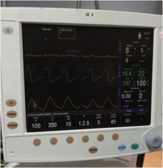
 Mechanical ventilation has long been an essential component of critical care medicine, saving countless lives in intensive care units (ICUs) and surgical settings. Pressure-Regulated Volume Control (PRVC), one of the several ventilatory modes that are currently accessible, has become a sophisticated choice that blends accuracy, flexibility, and patient safety. This blog explores the complexities of PRVC, its uses, and its importance, especially in view of current developments in medicine.

Image credit


What is PRVC Mode?

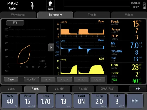
 The advantages of both volume-controlled and pressure-controlled ventilation are combined in the dual-control ventilation mode known as Pressure-Regulated Volume Control (PRVC). It is made especially to provide a predetermined tidal volume while automatically modifying inspiratory pressure to account for variations in the patient's airway resistance and lung compliance.


In PRVC, the ventilator continually analyzes the patient's respiratory mechanics and adjusts in real time to ensure adequate ventilation with the lowest risk of pressure-related lung damage. Because of this feature, PRVC is a vital tool for treating patients who are very sick.
Image Credit


How PRVC Works

  1. Set Tidal Volume: Clinicians set a desired tidal volume (amount of air delivered per breath) and a maximum pressure limit.

  2. Initial Breath: The ventilator delivers the first breath using an initial pressure estimate based on patient compliance.

  3. Feedback Mechanism: The ventilator measures the delivered tidal volume and adjusts the pressure for subsequent breaths to achieve the target volume.

  4. Continuous Adjustment: PRVC continuously recalibrates pressure within the set limits, providing consistent tidal volume delivery even as the patient’s lung mechanics change.

This adaptive mechanism ensures precise and safe ventilation, making PRVC an excellent choice for patients with variable lung conditions.

Why PRVC is a Game-Changer in Mechanical Ventilation

The unique ability of PRVC to combine the advantages of pressure control and volume control has redefined critical care ventilation. Below are some of the reasons why PRVC stands out:

1. Prevention of Barotrauma

One of the most significant risks in mechanical ventilation is barotrauma, caused by excessive airway pressures. PRVC minimizes this risk by dynamically adjusting inspiratory pressures to the lowest level required to achieve the set tidal volume.

2. Enhanced Patient-Ventilator Synchrony

Patient-ventilator dyssynchrony can lead to discomfort and prolonged ventilation. PRVC’s real-time adjustments promote better synchrony with the patient’s spontaneous breathing efforts, improving comfort and outcomes.

3. Optimized Oxygenation and Ventilation

PRVC ensures consistent delivery of tidal volume, which is crucial for maintaining adequate gas exchange in conditions like ARDS (Acute Respiratory Distress Syndrome).

4. Reduced Work of Breathing

For patients in respiratory distress, PRVC reduces the work required to breathe, allowing their lungs to rest and heal more effectively.

Image credit


Clinical Applications of PRVC

PRVC has proven invaluable across a range of clinical scenarios, including:

1. Management of ARDS

In ARDS, where lung compliance is severely compromised, PRVC provides precise ventilation by adjusting to rapidly changing pulmonary conditions, reducing the risk of ventilator-induced lung injury (VILI).

2. Pediatric and Neonatal Care

The delicate lungs of infants and children benefit from PRVC’s gentle approach, which ensures adequate ventilation without causing trauma.

3. Surgical Settings

During lengthy surgeries, lung compliance can fluctuate due to anesthesia and positioning. PRVC’s adaptive nature ensures consistent tidal volume delivery, enhancing intraoperative safety.

4. COVID-19 and Beyond

The COVID-19 pandemic placed unprecedented demands on mechanical ventilators. PRVC gained prominence for managing patients with COVID-19-induced ARDS, offering controlled ventilation while minimizing complications.

Considerations and Challenges

While PRVC offers numerous advantages, it is not without limitations. Clinicians must be aware of the following:

  1. Risk of Auto-PEEP: Patients with obstructive lung diseases may experience air trapping, leading to auto-positive end-expiratory pressure (auto-PEEP).

  2. Latency in Adjustments: Rapid changes in lung compliance may momentarily outpace the ventilator’s ability to adapt, potentially leading to under- or over-ventilation.

  3. Need for Expertise: PRVC requires careful monitoring and adjustments by trained clinicians to ensure optimal performance and patient safety.

A Vision for the Future

 PRVC and other ventilatory modes will likely grow even more advanced as technology develops. The design of ventilators is already incorporating advances in machine learning and artificial intelligence, which promise improved accuracy and patient-specific treatment. These developments are based on PRVC, which demonstrates the possibilities of intelligent, adaptive ventilation.

Why Understanding PRVC Matters

 Healthcare professionals can enhance patient outcomes and optimize resource use by having a thorough understanding of PRVC. Understanding ventilatory alternatives can help patients and their families communicate with care providers more effectively, enabling them to make well-informed decisions at crucial times.

Conclusion

 PRVC is a ground-breaking method that strikes a compromise between safety, flexibility, and accuracy in a world where mechanical ventilation frequently serves as the lifeline for critically ill patients. By seamlessly merging the strengths of pressure and volume management, PRVC not only addresses the complicated needs of critical care, but also paves the way for future advances in intelligent ventilatory support. Its contribution to improving patient care emphasizes how crucial ongoing innovation in medical technology.

Dec 25, 2024

Respiration Revolution: Mastering Ventilator Basics

Introduction

Despite being an essential component in modern medicine, ventilators can often remain mysterious to non-medical people. These devices are unsung heroes in the struggle against respiratory failure, helping patients with long-term respiratory conditions and even saving lives in intensive care units (ICUs). This blog will provide you with an interesting and simple explanation of how ventilators operate and why they are so important.


What is a Ventilator?

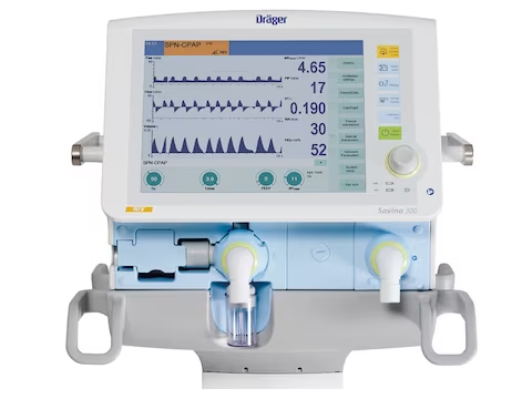
 Fundamentally, a ventilator is a device that assists or takes the place of breathing naturally. By ensuring a constant supply of oxygen to the body and the elimination of carbon dioxide, ventilators act like a bridge to recovery for people who are unable to breathe enough on their own because of disease, trauma, or surgery.


There are several different types of ventilators, that range from portable home appliances to advanced intensive care unit units. Maintaining proper gas exchange in the lungs is their fundamental objective, regardless of the setting.
Image Credit


How Does a Ventilator Work?

Ventilators work by delivering pressurized air or a mixture of air and oxygen through a tube placed in the patient’s airway (invasive ventilation) or via a mask (non-invasive ventilation). They operate based on key principles:

  • Pressure: Ensures that the lungs inflate and deflate effectively.

  • Volume: Delivers a specific amount of air with each breath.

  • Rate: Controls how many breaths the ventilator delivers per minute.

These variables can be adjusted depending on the patient’s condition and needs, making ventilators versatile tools in medical care.

Why Are Ventilators So Important?

Ventilators are more than just machines; they are lifelines. During the COVID-19 pandemic, they became symbols of hope, as they were vital for patients suffering from severe respiratory distress. Beyond emergencies, ventilators play a key role in managing chronic conditions such as:

  • Chronic obstructive pulmonary disease (COPD)

  • Neuromuscular disorders like ALS

  • Post-surgical recovery

For healthcare providers, ventilators are invaluable in saving lives and giving patients a fighting chance.

Image Credit


A Glimpse into Ventilator Modes

Ventilator modes determine how the machine supports breathing. While we’ll dive deeper into this topic in upcoming blogs, here’s a teaser:

  • Assist-Control (AC): A mode where the ventilator provides full support with each breath.

  • Synchronized Intermittent Mandatory Ventilation (SIMV): Balances machine-delivered and spontaneous breaths.

  • Continuous Positive Airway Pressure (CPAP): Maintains airway pressure without initiating breaths.

Stay tuned for an in-depth exploration of these modes and more!

Dec 24, 2024

Unmasking the Silence: Exploring Apnea Testing

 Introduction

The irreversible cessation of all brain activity, including that of the brainstem and cerebral cortex, is known as brain death. In clinical settings, determining brain death is a crucial process, particularly when making decisions about organ donation and life support withdrawal. This blog talks about the Doll's Eye Reflex, Cold Caloric Test, Gag Reflex, and Vestibular Reflex—the four main tests used to evaluate brainstem function.

1. The Doll's Eye Reflex (Oculocephalic Reflex)

The Doll's Eye reflex is an oculocephalic response that assesses brainstem function. This test is based on the idea that if the brainstem is working properly, the eyes should to turn in the opposite direction when the head is turned.

  • Procedure:

    1. The patient's eyelids are gently opened.

    2. The examiner rapidly but carefully rotates the head from side to side or flexes it up and down.

  • Normal Response:  The eyes move opposite to the direction of head movement. For example, if the head is turned to the right, the eyes move to the left.

  • Abnormal Response: A lack of eye movement indicates a non-functional brainstem. This test cannot be performed in cases of cervical spine instability.

  • Clinical Significance: The absence of the Doll’s Eye reflex suggests brainstem dysfunction, supporting a diagnosis of brain death when combined with other tests.
Image credit


2. Cold Caloric Test (Oculovestibular Reflex)

The Cold Caloric Test assesses the vestibulo-ocular reflex, which depends on an intact brainstem.

  • Procedure:

    1. Ensure the patient's tympanic membrane is intact.

    2. Elevate the head to 30 degrees to align the horizontal semicircular canals.

    3. Instill approximately 50 mL of cold water (0–5°C) into one ear canal using a syringe.

  • Normal Response: The eyes will deviate toward the irrigated ear, followed by a nystagmus (rapid movement) in the opposite direction. This response indicates an intact brainstem.

  • Abnormal Response: No eye movement occurs in patients with brainstem death. Absence of this reflex is a critical indicator.

  • Considerations: Ensure no ear pathology that might affect the results. This test can cause discomfort, so it’s only performed in unresponsive patients.

3. Gag Reflex (Pharyngeal Reflex)

The gag reflex is a brainstem-mediated response to stimulation of the posterior pharyngeal wall.

  • Procedure:

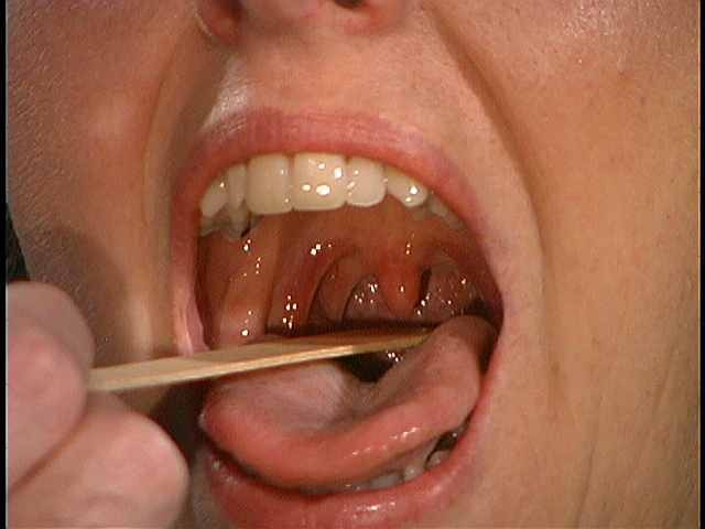
    1. Using a sterile swab or tongue depressor, gently stimulate the posterior pharynx or soft palate.

  • Normal Response: Contraction of the pharyngeal muscles causing gagging or retching.

  • Abnormal Response: Absence of a gag reflex indicates dysfunction of cranial nerves IX (glossopharyngeal) and X (vagus), which are coordinated in the brainstem.

  • Clinical Significance: Loss of the gag reflex, when accompanied by other absent brainstem reflexes, is a significant indicator of brain death.

4. Vestibular Reflex (Vestibulo-Ocular Reflex)

The vestibular reflex maintains gaze stability by coordinating eye movements with head movements.

  • Assessment in Brain Death Testing:

    1. This reflex is indirectly assessed through tests like the Doll’s Eye reflex and the Cold Caloric Test.

    2. Absence of eye movements in both tests confirms the loss of vestibulo-ocular reflexes.

  • Clinical Insight: A functional vestibulo-ocular reflex signifies intact connections between the inner ear, brainstem, and extraocular muscles.

Conclusion

The Doll’s Eye Reflex, Cold Caloric Test, Gag Reflex, and Vestibular Reflex are cornerstone assessments in the determination of brain death. Each test targets specific components of brainstem function, ensuring a comprehensive evaluation. Brain death testing is meticulously standardized to exclude confounding factors like drug intoxication, hypothermia, or metabolic disturbances. These tests, combined with ancillary studies like EEG or cerebral blood flow imaging, provide a robust framework for confirming brain death.

Dec 23, 2024

Breathing Through History: The Legacy of Negative Pressure Ventilation and the Iron Lung

Introduction

In the development of respiratory care, negative pressure ventilation (NPV) has been essential, and its most recognizable emblem is the iron lung. With a long history, this technology replicates the natural breathing mechanics and still has an impact on modern medical practices.

Image credit


The Mechanics of Negative Pressure Ventilation

The diaphragm contracts during natural respiration, drawing air into the lungs through a vacuum. By enclosing the patient's body—apart from the head—in a sealed chamber, NPV mimics this process. Air can enter the lungs by expanding the chest cavity and decrease the pressure inside the chamber. When the pressure returns to normal, the chest contracts and expels air. In contrast, positive pressure ventilation uses intubation to drive air directly into the lungs.

The Iron Lung: A Lifesaving Apparatus

 The iron lung, which was designed in the late 1920s, came to represent the struggle against polio, a disease that often paralyzed respiratory muscles. Patients would lie in the iron lung for extended periods of time, with only their heads exposed. The machine's periodic pressure changes make breathing easier, providing a lifeline to many who might otherwise succumbed of respiratory failure.

Image credit


Modern Perspectives and Continued Use

 Iron lungs have mostly been replaced by positive pressure ventilators because of their portability and user-friendliness, but NPV is still relevant. Notably, it offers a breathing rhythm that is more physiological and may be beneficial in some conditions. For example, NPV devices gained attention again during the COVID-19 epidemic as potential substitutes for traditional ventilators.


Some people who survived polio still utilize iron lungs, which is remarkable. Their stories demonstrate the technology's long-term viability and great impact on their lives. For example, Paul Alexander, who contracted polio in 1952, relied on an iron lung for more than 60 years.
Image credit


Advancements in Respiratory Care

Modern ventilators are more effective and patient-friendly thanks to the legacy of the iron lung. Today's technologies provide complex ways of ventilation that are tuned to individual patient demands, and they have become essential tools in intensive care units worldwide.

Conclusion

The evolution of medical technology from the iron lung to modern ventilators demonstrates its inventiveness and adaptability. Understanding the history and mechanics of negative pressure ventilation respects prior accomplishments while also inspiring future advances in respiratory care.

Dec 22, 2024

Unraveling Mpox: A Modern Challenge in Global Health

 Unraveling Mpox: A Modern Challenge in Global Health


Emerging disease outbreaks have frequently taken center stage in the recent problems facing the global health landscape. Mpox, formerly known as monkeypox, is one such disease. Despite being around for decades, outbreaks outside of its natural endemic regions have made it more significant in terms of public health. The purpose of this blog is to provide information about Mpox, its effects on human health, and the steps we can take to safeguard our communities and ourselves.


 What is Mpox?

 The Mpox virus, which relates to the Orthopoxvirus genus, is the cause of the viral infection known as mpox. Although Mpox is typically less severe, it is closely related to the virus that causes smallpox. Although rodents are probably the disease's natural hosts, it was initially discovered in 1958 in study monkeys, hence its original name. The Democratic Republic of the Congo reported the first human case in 1970.

Symptoms and Transmission

 Flu-like symptoms like fever, chills, and muscular aches are the first signs of mpox. A rash that usually starts on the face and spreads to other areas of the body follows. After going through several phases, the rash finally forms scabs and falls off.


Close contact with an infected person or animal, as well as contaminated bedding or other materials, can spread the infection. Human-to-human transmission mostly occurs by bodily fluids, direct contact with lesions, or respiration droplets. For prevention, it is essential to comprehend these ways of transmission.

Why Mpox Matters Today

 For many years, Mpox was limited to specific areas in Central and West Africa. However, recent outbreaks in countries where disease is not endemic caused  around the world. Factors such as increased international trips, climatic change, and low smallpox vaccination coverage—which provides some cross-protection against Mpox—may all contribute to its spread.


The 2022 outbreak, for example, included patients from Europe, North America, and other countries, indicating how linked our globe has grown. The necessity of strong surveillance systems and quick response plans was highlighted by this outbreak.

Prevention and Protection

 Prevention depends on practical steps and public awareness:
Vaccination: It has been demonstrated that the smallpox vaccine is effective against Mpox. Vaccination initiatives can aid in controlling the spread of an outbreak.

Hygiene Practices: It's crucial to wash your hands frequently and keep your distance from infected people or animals.

Community Engagement: Encouraging open communication and combating disinformation can help communities act responsibly.

Healthcare Access: Timely diagnosis, treatment, and containment are ensured by strengthening healthcare systems.

The Human Angle

 Beyond the science, it is critical to understand the human stories around Mpox. Outbreaks frequently disproportionately impact underprivileged areas with limited access to healthcare. The stigma associated with the illness can make it more difficult for people to report cases and seek treatment, which will accelerate its spread. Compassionate communication and inclusive policy are critical for tackling these issues.

Looking Ahead

 As the globe continues to struggle with Mpox, collaboration is our most effective tool. Governments, healthcare professionals, researchers, and individuals must collaborate to create robust systems capable of combating not only Mpox, but also other emerging infectious diseases.

Dec 21, 2024

Threads of Immunity: Unraveling Wiskott-Aldrich Syndrome

 Threads of Immunity: Unraveling Wiskott-Aldrich Syndrome


Introduction

Wiskott-Aldrich Syndrome (WAS) is a rare genetic disorder that predominantly affects males and is characterized by a triad of symptoms: eczema, thrombocytopenia (low platelet count), and recurrent infections. This condition is caused by mutations in the WAS gene, which encodes the Wiskott-Aldrich Syndrome protein (WASP), crucial for the proper functioning of blood cells and immune responses.


What Causes Wiskott-Aldrich Syndrome?

WAS is an X-linked recessive disorder, meaning the defective gene is located on the X chromosome. Since males have only one X chromosome, a single mutation in the WAS gene can lead to the disease. Females, having two X chromosomes, are typically carriers and usually do not exhibit symptoms, although in rare cases, they may have mild manifestations.

The WASP protein plays a critical role in the cytoskeleton of blood cells, which is essential for cell signaling, movement, and interaction. Mutations in the WAS gene disrupt these processes, leading to the clinical features of the disease.

Eczematous lesions in Wiskott-Aldrich syndrome. The lesion is essentially indistinguishable from that of atopic dermatitis except for the presence of purpura and petechiae.


Key Symptoms of Wiskott-Aldrich Syndrome

  1. Thrombocytopenia:

    • Low platelet count leads to easy bruising, prolonged bleeding, and the presence of small platelets on blood tests.

  2. Eczema:

    • Patients often suffer from persistent and severe eczema, a chronic skin condition characterized by redness, itching, and inflammation.

  3. Recurrent Infections:

    • Due to immune system dysfunction, individuals are prone to bacterial, viral, and fungal infections, often severe or life-threatening.

  4. Autoimmune Disorders and Cancers:

    • Some patients develop autoimmune diseases such as hemolytic anemia or vasculitis. Additionally, there is an increased risk of lymphomas and other cancers.


Diagnosis

Diagnosis of WAS typically involves:

  • Clinical Evaluation: Based on characteristic symptoms.

  • Blood Tests: To assess platelet count, size, and immune function.

  • Genetic Testing: To confirm mutations in the WAS gene.

Prenatal testing is also available for families with a known history of the condition.

Treatment Options

  1. Hematopoietic Stem Cell Transplantation (HSCT):

    • HSCT is the only curative treatment for WAS. Early transplantation significantly improves outcomes.

  2. Supportive Care:

    • Includes platelet transfusions, immunoglobulin replacement therapy, and antibiotics to manage symptoms and prevent infections.

  3. Gene Therapy:

    • An emerging treatment that involves correcting the defective gene in the patient’s stem cells. Clinical trials have shown promise.

  4. Medications:

    • Immunosuppressive drugs may be used for autoimmune complications.

Living with Wiskott-Aldrich Syndrome

Managing WAS requires a multidisciplinary approach involving hematologists, immunologists, and dermatologists. Families should be educated about infection prevention, the importance of regular medical follow-ups, and the potential need for genetic counseling.

Recent Advances in Research

Research efforts are focused on understanding the molecular mechanisms of WAS and improving treatment options. Gene therapy is particularly promising, offering hope for a long-term cure without the need for donor stem cells.

Conclusion

Wiskott-Aldrich Syndrome is a complex disorder with significant challenges for affected individuals and their families. Advances in genetic testing, supportive care, and innovative therapies have improved the prognosis for many patients. Early diagnosis and a comprehensive treatment approach are essential for better outcomes.

Dec 20, 2024

THE DANCING VIRUS

THE DANCING VIRUS[DINGA-DINGA]

In December 2024, a mysterious illness known as "Dinga Dinga" emerged in Uganda's Bundibugyo district, predominantly affecting women and girls.The term "Dinga Dinga," translating to "shaking like dancing," aptly describes the condition's hallmark symptom: uncontrollable body shaking that impedes mobility. 

Symptoms and Impact

Individuals afflicted with Dinga Dinga experience:

  • Uncontrollable Shaking: Intense, involuntary movements resembling dance-like motions.

  • Fever and Fatigue: High fever accompanied by extreme weakness.

  • Mobility Challenges: The severity of symptoms often renders walking difficult or nearly impossible.

Approximately 300 cases have been reported, with no fatalities to date.Most patients recover within a week following appropriate medical treatment. 

Health Authorities' Response

Local health officials are actively investigating the cause of Dinga Dinga.Samples from affected individuals have been sent to Uganda's Ministry of Health for analysis.In the interim, treatment with antibiotics has proven effective, leading to patient recovery within a week.Dr. Kiyita Christopher, the District Health Officer, advises against the use of unverified herbal remedies and urges those experiencing symptoms to seek care at local health facilities. 

Historical Context

 The sudden onset and distinctive symptoms of Dinga Dinga have drawn comparisons to historical phenomena such as the "Dancing Plague" of 1518 in Strasbourg, France, where individuals danced uncontrollably for days.However, any direct correlation remains speculative pending further investigation. 

Current Status and Recommendations

As of now, Dinga Dinga cases are confined to the Bundibugyo district, with no reports from other regions.Health authorities continue to monitor the situation closely, emphasizing the importance of early medical intervention.Residents are encouraged to remain vigilant and to seek prompt medical attention if symptoms manifest. 

In summary, while Dinga Dinga presents with alarming symptoms, the high recovery rate with appropriate treatment offers reassurance.Ongoing investigations aim to uncover the underlying cause of this enigmatic illness to inform future prevention and treatment strategies.

Dec 16, 2024

Whispers of the Laughing Death: The Mystery of Kuru

Whispers of the Laughing Death: The Mystery of Kuru


Introduction 

Kuru disease is a rare and fatal neurodegenerative disorder that has intrigued scientists and healthcare professionals for decades. Originating among the Fore people of Papua New Guinea, this disease is part of a group of conditions known as transmissible spongiform encephalopathies (TSEs), which include Creutzfeldt-Jakob disease and mad cow disease. This blog will delve into the origins, symptoms, causes, prevalence, and current understanding of Kuru, a disease that provides unique insights into human biology and cultural practices.


What is Kuru Disease?

Kuru, which means "to shiver" or "tremble" in the Fore language, derives its name from the tremors and motor dysfunction that characterize the disease. It primarily affects the central nervous system, leading to progressive neurological decline and, eventually, death. The disease is caused by prions—misfolded proteins that trigger a cascade of abnormal folding in normal brain proteins, resulting in brain tissue damage.

Origins and Cultural Context

Kuru was first documented in the mid-20th century among the Fore people of Papua New Guinea. It was discovered that the disease was closely linked to a unique cultural practice: ritualistic cannibalism. As part of mourning rituals, the Fore consumed the bodies of deceased relatives, believing this act helped free the spirits of the dead. Women and children, who were more likely to consume brain tissue—the part of the body most heavily laden with infectious prions—were disproportionately affected by Kuru.


Symptoms and Progression

Kuru progresses through three distinct stages:

  1. Ambulant Stage: Early symptoms include unsteady gait, tremors, and slurred speech. Patients may also experience subtle behavioral changes and difficulty in coordination.

  2. Sedentary Stage: As the disease progresses, individuals become unable to walk, experience severe tremors, muscle jerks (myoclonus), and emotional instability.

  3. Terminal Stage: In the final stage, patients are bedridden and develop severe neurological symptoms, including incontinence, dysphagia (difficulty swallowing), and unresponsiveness. Death usually occurs within 12 months of symptom onset due to complications such as pneumonia.

Cause and Mechanism

Prions, the infectious agents responsible for Kuru, are abnormal forms of the prion protein (PrP). Unlike viruses or bacteria, prions lack genetic material and are resistant to standard methods of sterilization. When these misfolded proteins enter the brain, they induce normal prion proteins to adopt the same abnormal structure, leading to a chain reaction that results in extensive brain damage. The spongy appearance of affected brain tissue, observed under a microscope, is characteristic of TSEs.


Prevalence and Decline

Kuru reached its peak in the 1950s and 1960s, with over 1,000 deaths reported annually among the Fore people. The disease has since nearly disappeared following the cessation of ritualistic cannibalism, thanks to public health interventions and cultural shifts. The last known cases of Kuru were reported in the late 2000s, attributed to the long incubation period of the disease, which can exceed 30 years.

Scientific Significance

Kuru has provided valuable insights into prion diseases and human genetics. Researchers have studied the Fore population to understand genetic resistance to prion diseases. For instance, a protective mutation in the PRNP gene has been identified in some individuals, offering clues for potential therapies for other prion-related disorders.


Conclusion

Kuru disease is a poignant reminder of the intricate interplay between culture, biology, and disease. Although this devastating condition has been virtually eradicated, its legacy lives on in scientific research and our understanding of prion diseases. By studying Kuru, we not only honor the lives lost but also advance our knowledge of human health and resilience.

Dec 14, 2024

Cracking the Code of DLCO: Unveiling Lung Health

Cracking the Code of DLCO: Unveiling Lung Health


INTRODUCTION

When assessing lung function, one of the key tests often performed is the diffusing capacity of the lungs for carbon monoxide (DLCO). This test offers valuable insights into how well the lungs are facilitating gas exchange — a critical function for oxygen delivery to the body. In this blog, we will explore the science behind DLCO, its clinical relevance, how it is measured, and what the results mean for patients and healthcare providers.

What Is DLCO?

DLCO measures how efficiently oxygen and carbon dioxide are exchanged between the alveoli (tiny air sacs in the lungs) and the blood. By using carbon monoxide (CO) as a marker, this test evaluates the ability of the lungs to transfer gases because CO binds to hemoglobin in a way similar to oxygen, but with much greater affinity.

Carbon monoxide is chosen for its high binding efficiency and because it occurs in negligible amounts in the body under normal conditions, making it an ideal tracer for gas exchange studies.

Why Is DLCO Important?

DLCO provides critical information in diagnosing and managing a range of pulmonary conditions. These include:

  1. Obstructive lung diseases: Conditions like chronic obstructive pulmonary disease (COPD) and asthma can reduce DLCO due to airway obstructions and damage to alveoli.

  2. Restrictive lung diseases: Interstitial lung diseases (ILD), pulmonary fibrosis, and sarcoidosis can impair DLCO by thickening or scarring the alveolar membrane.

  3. Pulmonary vascular diseases: Conditions such as pulmonary hypertension or chronic thromboembolic disease may decrease DLCO by reducing blood flow in the pulmonary capillaries.

  4. Other systemic diseases: Autoimmune diseases like systemic lupus erythematosus and scleroderma often manifest with lung involvement, and a reduced DLCO can be an early indicator of pulmonary complications.

How Is DLCO Measured?

The DLCO test is performed using a spirometer and involves the following steps:

  1. Inhalation: The patient breathes in a gas mixture containing a trace amount of carbon monoxide and an inert gas (such as helium).

  2. Breath-holding: The patient holds their breath for approximately 10 seconds, allowing gas exchange to occur in the lungs.

  3. Exhalation: The exhaled gas is analyzed to determine the concentrations of carbon monoxide and inert gas.

From this data, the diffusing capacity is calculated, typically adjusted for hemoglobin levels and lung volume to ensure accuracy.

Interpreting DLCO Results

DLCO results are typically reported as a percentage of the predicted value based on age, sex, height, and ethnicity. Results are categorized as:

  • Normal DLCO: Typically 80-120% of the predicted value, indicating efficient gas exchange.

  • Low DLCO: Below 80% of the predicted value, suggesting impaired gas exchange. This is commonly seen in conditions like pulmonary fibrosis or emphysema.

  • High DLCO: Above 120% of the predicted value, less common but can occur in cases of polycythemia or alveolar hemorrhage.

Parameters and graphs of DLCO determination. DLCO – 81% of predicted, normal range; VA – alveolar vol-ume 73% of predicted, a decreased parameter


Factors Influencing DLCO

Several physiological and pathological factors can affect DLCO:

  • Hemoglobin levels: Anemia reduces DLCO, while polycythemia increases it.

  • Body positioning: DLCO is higher in the supine position compared to standing.

  • Smoking: Smokers may have reduced DLCO due to alveolar damage and the presence of carbon monoxide in their blood.

  • Altitude: Living at higher altitudes can increase DLCO due to physiological adaptations.

Clinical Application of DLCO

DLCO results are often used in conjunction with other pulmonary function tests (PFTs) to provide a comprehensive view of lung health. For example:

  • Distinguishing diseases: In COPD, DLCO is usually reduced in emphysema but normal in chronic bronchitis. Similarly, in asthma, DLCO is typically normal or increased.

  • Monitoring disease progression: In ILD or pulmonary hypertension, a declining DLCO may indicate worsening disease.

  • Preoperative assessment: Patients undergoing major surgeries, especially lung resections, may undergo DLCO testing to assess their ability to tolerate reduced lung function.

Improving DLCO

In some cases, improving DLCO is possible by addressing the underlying condition. For example:

  • Quitting smoking: Reduces ongoing lung damage and improves gas exchange over time.

  • Managing anemia: Correcting low hemoglobin levels can improve DLCO.

  • Treating primary disease: Effective management of pulmonary conditions such as interstitial lung disease or pulmonary hypertension can stabilize or even improve DLCO.

Conclusion

DLCO is a powerful tool in the arsenal of respiratory diagnostics, helping healthcare providers understand and manage a variety of lung conditions. Whether you are a patient looking to understand your test results or a healthcare professional seeking to deepen your knowledge, understanding DLCO is crucial for informed decision-making.

Dec 13, 2024

Breath of Insight: Exploring Spirometry and Body Plethysmography

 Breath of Insight: Exploring Spirometry and Body Plethysmography


Introduction

Pulmonary function tests (PFTs) are essential tools in diagnosing and monitoring respiratory diseases. Among the most widely used PFTs are spirometry and body plethysmography. These tests provide valuable insights into lung function and help clinicians assess conditions such as asthma, chronic obstructive pulmonary disease (COPD), and restrictive lung diseases. This blog delves into the principles, procedures, and clinical significance of spirometry and body plethysmography, supported by recent advancements and research.

What is Spirometry?

Spirometry is a non-invasive test that measures the volume and flow of air that a person can inhale and exhale. It provides critical data on lung capacity and airflow limitation, which are key indicators of respiratory health.

How Does Spirometry Work?

During spirometry, the patient breathes into a device called a spirometer. The test typically involves:

  1. Forced Vital Capacity (FVC): The total volume of air exhaled forcefully after taking a deep breath.

  2. Forced Expiratory Volume in 1 Second (FEV1): The volume of air exhaled in the first second of the FVC maneuver.

  3. FEV1/FVC Ratio: A critical parameter used to identify obstructive or restrictive lung patterns.


 (A) Flow-volume curve. (B) Volume-time curve. The smooth lines, expiratory time of greater than six seconds, and quick peak of the peak expiratory flow rate indicate a good spirometric effort.


Clinical Applications of Spirometry

  • Obstructive Lung Diseases: Conditions like asthma and COPD often show a reduced FEV1/FVC ratio.

  • Restrictive Lung Diseases: Characterized by reduced FVC but a normal or increased FEV1/FVC ratio.

  • Screening and Monitoring: Regular spirometry is vital for tracking disease progression and response to treatment.

Advancements in Spirometry

Recent innovations include portable and digital spirometers that provide real-time feedback and data integration with electronic health records (EHRs). These advancements enhance diagnostic accuracy and patient accessibility.

What is Body Plethysmography?

Body plethysmography is a more comprehensive test that measures lung volumes and airway resistance. It is particularly useful when spirometry cannot provide conclusive results.


How Does Body Plethysmography Work?

The test is conducted in an airtight chamber where the patient breathes through a mouthpiece. By measuring pressure changes in the chamber and the airway, clinicians can calculate:

  • Total Lung Capacity (TLC): The maximum volume of air in the lungs after a full inhalation.

  • Residual Volume (RV): The volume of air remaining in the lungs after maximal exhalation.

  • Airway Resistance (Raw): The resistance to airflow within the respiratory tract.

Clinical Applications of Body Plethysmography

  • Restrictive Lung Diseases: Differentiates between intrinsic and extrinsic restrictive patterns by assessing lung volumes.

  • Obstructive Lung Diseases: Provides precise measurements of air trapping and hyperinflation in conditions like COPD.

  • Preoperative Assessments: Evaluates lung function before thoracic or abdominal surgeries.

Advancements in Body Plethysmography

Modern plethysmographs feature advanced sensors and software that improve the accuracy and speed of measurements. Integration with imaging technologies, such as CT scans, offers a more holistic view of lung function and structure.

Comparing Spirometry and Body Plethysmography


Feature

Spirometry

Body Plethysmography

Purpose

Measures airflow and lung capacity

Measures lung volumes and airway resistance

Accuracy

Limited for lung volumes

Highly accurate for lung volumes

Clinical Use

Initial screening and monitoring

Detailed diagnosis and advanced assessment

Equipment

Portable spirometers

Specialized airtight chambers


Conclusion

Both spirometry and body plethysmography are indispensable in the evaluation of respiratory health. While spirometry serves as a frontline diagnostic tool, body plethysmography provides detailed insights that complement initial findings. With advancements in technology, these tests are becoming more accessible, accurate, and integral to personalized respiratory care.

Turning Back Time on Cancer|Korean Scientists Discover Reprogramming Breakthrough

Introduction The quest to treat cancer effectively and with minimal side effects has taken a monumental leap forward, thanks to Korean scien...